Keep the Body's First Line of Defense Strong

Assess Your Mucosal Immune System with Array 14

Immune Tolerance/Immune Resilience

We are all constantly exposed to chemicals, foods, viruses and pathogens that can do us harm.  Immune tolerance provides our immune system the ability to react to environmental triggers.  Therefore, when you have proper immune tolerance, your body defense mechanisms protect you from chemical reactivity, food reactivity and autoimmune reactivity.  Immune tolerance includes “oral tolerance.”  This process allows your body to tolerate commonly consumed foods.  When you lose immune tolerance, food sensitivities can develop.

Chemical Intolerance

Chemical intolerance is another type of immune intolerance.  If you have lost chemical tolerance, your body can react to personal products such as perfumes, lotions, shampoos, soaps, household cleaning products, and detergents, just to name a few.  Frequent exposure to these exogenous substances can break down your first line of defense, the mucosal immune system.

Autoimmune Reactivity

Loss of “self” tolerance causes the body to abnormally attack itself, resulting in autoimmune reactivity.  Therefore, it is important to understand that loss of immune tolerance can affect how resilient you are to your environment.

Immune Resilience

Immune resilience is the capacity of the immune system to return to a healthy state of well-being, known as homeostasis.  When your immune system is balanced, you maintain the ability to fight off pathogens and stress, and to adapt to adverse environmental exposures.  Chronic immune activation by  viruses and bacteria promotes loss of tolerance, and subsequent dysregulation of our immune system.

Improving immune resilience is the key to supporting immune tolerance.  Preventing the breakdown of immune system tolerance is largely influenced by the strength of the immune barriers.

The Mucosal Immune System is The First Line of Defense

The mucous membrane is the body’s first line of immune system defense, which builds a protective wall against antigen infiltration and protects the body from environmental attacks. This important barrier intercepts viruses, food proteins and peptides, chemicals, and bacteria. The membrane lines and protects various cavities in the body and covers the surface of internal organs.

The Immune System’s Defensive wall is composed of a mucous layer that contains secretory immunoglobulin A (IgA) antibodies. SIgA antibodies seize antigenic invaders before they can penetrate epithelial cells and eliminate them through the digestive track.

Assessing the First Line of Defense

It is critical to assess the first line of defense. A weakened immune system can lead to a vulnerability to viruses, food proteins and peptides, airborne molecules and bacterial antigens, placing the body at greater risk for the development of autoimmunity.

Immunoglobulin A (IgA) is an antibody playing a critical role in mucosal immunity.  IgA accounts for approximately 15% of serum immunoglobulins, but it is most abundant in saliva, tears, and in the secretions of the esophagus, lungs, and gastrointestinal tract.  In secretory form, IgA (sIgA) is the main immunoglobulin found in mucous secretions.  This mucosal antibody provides protection due to its ability to survive in harsh environments like the respiratory and gastrointestinal tract.

What is the role of Secretory IgA? SIgA is a major component of the defensive wall which serves to function as the first responder and prevent hostile viruses, pathogens, and other environmental factors from breaking through the body’s defenses. This mucosal immune barrier functions like Velcro™, blocking the infiltrator and stopping it from binding to the epithelial receptors.

In saliva and gastrointestinal secretions, SIgA is the antibody that reacts to these invaders and prevents them from damaging the gastrointestinal system.

IgA deficiency and autoimmunity

SIgA deficiency and autoimmunity. Secretory IgA in the Fc-a Receptor1 acts as a blocker and inhibits cell activation that could lead to immune dysregulation and autoimmunity. An empty FcaR1 with no SIgA means no inhibition of cell activation, resulting in autoimmunity. In individuals with IgA deficiency, IgM can compensate by assuming IgA’s functions and playing a similar protective role.

Mucosal Tolerance

Salivary Testing

Many patients with food sensitivity conditions, including non-celiac gluten sensitivity, celiac disease, parasitic infection, gut dysbiosis and inflammatory bowel disease, leave identifiable and specific IgA and IgM immune imprints in saliva. Repeated exposure to a reactive antigen in the mucosa that crosses the intestinal barriers can result in excessive antibody production in the blood. Saliva is a source of body fluid for detection of immune antibody responses (90% IgA and 10% IgM).

The Benefits of Salivary Testing

  • Salivary testing offers insight into the world of mucosal immunity, indicating possible failure of oral tolerance and possible immune dysregulation in the gut.
  • Saliva contains antibodies against bacterial, viral, parasitic, food and tissue antigens, and enzymes present in the oral cavity and gastrointestinal tract. Saliva testing can reveal exposure and immune reaction to these antigens.
  • A salivary specimen is ideal for the detection of gastrointestinal inflammation and possible autoimmunity at the earliest stage, before the appearance of antibodies in the blood.
  • Oral fluid collection is also a good alternative for antibody measurement when a serum collection is not possible.
  • Saliva collection is non-invasive, painless, and convenient for the patient.

Dr. Chad Larson talks about SARS-2

Cyrex Testing – Array 14 Mucosal Immune Reactivity Screen™

A comprehensive, breakthrough test that gives you the big picture by encompassing in the Cyrex System™

Essential Barrier Permeability

Environmental Triggers

Predictive Antibodies of Autoimmune Reactivity

The panel tests selected antigens for salivary IgA+IgM antibody measurement: Secretory IgA; gut tight junctions and structural proteins that are involved in inflammation and gut dysbiosis; most commonly reactive foods; toxic chemicals; pathogens; and blood-brain barrier integrity markers.

Clinical Use:

  • Evaluate mechanisms of compromised immune tolerance
  • Evaluate possible outcomes of compromised mucosal tolerance, such as: intestinal barrier dysfunction, food and chemical immune reactivity, and autoimmunity
Recommended for Patients Who:

  • Have chronic inflammatory or irritable bowel conditions
  • Have a family history of autoimmune disease
  • Have malabsorption or maldigestion
  • Have had antibiotic overuse
  • Have intestinal bacteria or yeast overgrowth
  • Have intestinal infections
  • Have gastroenteritis
  • Have had food poisoning (loss of tolerance)

Frequently Asked Questions

The mucous membrane is a membrane that lines various cavities in the body and covers the surface of internal organs. It consists of one or more layers of epithelial cells overlaying a layer of loose connective tissue (lamina propria). It protects various body openings such as the eyes, ears, the insides of the mouth and the nose, the reproductive tract, and internal organs such as the gastrointestinal (GI) tract. Due to its large surface, the GI tract is exposed to a significant amount of intestinal contents, including food, bacteria and their toxins. It is a major entry point for pathogens, bacterial toxins, toxic chemicals and undigested food antigens. The gut mucosa is known to be the largest immunological environment of the body.

Although it may vary slightly from location or organ to organ, the mucous membrane generally has a protective mucus layer that contains secretory IgA (SIgA), antibodies that comprise the first line of antigen-specific immune defense. The mucus layer acts like velcro, catching and stopping pathogens, antigens and allergens from having contact with the epithelial cell layer and promoting their clearance into and through the waste system.

This clearance of unwanted cells and molecules by the GI mucus layer not only prevents the binding of pathogens or antigens to the epithelial cells but also promotes the inhibition of inflammation and autoimmunity.

In the absence of the mucous membrane, bacteria, viruses, parasites, yeasts, the various toxins of the foregoing, allergens, toxic chemicals and food components can penetrate the gut barriers and enter into the submucosa, regional lymph nodes and circulation. The entry of antigens into the circulation and tissues, activates the innate and adaptive immune responses; the ensuing antibody production against these antigens may result in inflammation and autoimmune reaction.

The functions of the mucous membrane are to prevent the entry of pathogens, and antigens into the body, to protect the body from being infected by viruses and bacteria, and to keep the mucosal tissues adequately moisturized so that the pathogens in viral droplets will not be able to bind to specific receptors on epithelial cells and penetrate their structures.

Saliva is a mucosal body fluid with important functions in oral, gastro-intestinal and general health. The three major salivary glands, parotid, submandibular, and sublingual, provide more than 90% of salivary secretion. The rest comes from hundreds of minor salivary glands distributed throughout the oral cavity. Saliva helps clean the mouth, digest food, balance pH, and fight tooth decay. It also carries many of the same proteins and other molecules, including antibodies, that are found in blood and urine.

Secretory immunoglobulin A (SIgA) is a very unique antibody that is a major component of the mucosal defensive wall against environmental antigens with which the mucosal immune system comes in contact. In saliva and gastrointestinal secretions, SIgA is the antibody that reacts against various antigens and prevents them from damaging the GI system. Figure 1 shows how the lack of SIgA due to IgA deficiency could lead to autoimmunity. In individuals with IgA deficiency, IgM compensates by assuming IgA’s functions and playing the same protective role.

Figure 1. IgA deficiency and autoimmunity. Secretory IgA in the Fc-α receptor1 acts as a blocker and inhibits cell activation that could lead to autoimmunity. An empty Fc-α receptor1 with no SIgA means no inhibition of cell activation, resulting in autoimmunity.

  • IgA and IgM antibody production against various antigens in saliva may be an indication of a defect in oral tolerance which can result in inflammatory conditions in the bowel. Mucosal or oral tolerance is the suppression or downregulation of immune cell responses to an antigen by prior exposure of the antigen through the mucosal route. Defects in the mechanism of oral tolerance have been reported as being responsible for several diseases of the gastrointestinal and respiratory tract, in particular, gastric autoimmunity.
  • In fact, patients with food sensitivity conditions, including non-celiac gluten sensitivity, celiac disease, parasitic infection, gut dysbiosis, and inflammatory bowel disease leave identifiable and specific IgA and IgM immune prints in saliva. However, repeated exposure to an antigen in the mucosa that crosses the intestinal barriers results in antibody production in the blood. Therefore, saliva antibodies (SigA) may be detecting an immune reaction to antigens in their initial exposure stage, in which they may not yet have gotten into the circulation.
  • Based on this mechanism of action, saliva is an excellent body fluid for use in the detection of an immune response (90% IgA and 10% IgM) to antigens present in the oral cavity and gastrointestinal tract. Indeed, salivary antibody induction has been widely used as a model system to study secretory responses to ingested material, primarily because saliva is easy to collect and analyze.
  • Since the mucosa is the first line of defense, elevated salivary antibodies to the gut microbiota, gut-associated antigens, dietary proteins, xenobiotics and enteric nervous system (ENS) antigens assessed in Array 14 may be a warning of mucosal immune over-reactivity against these antigens. If this specific mucosal immune reactivity is not addressed, the intestinal barrier may be breached by the environmental insult. This would make it possible for environmental toxins to infiltrate the body, and the resulting inflammation may eventually lead to the onset of autoimmune or neuroautoimmune disorders.
  • Salivary testing offers insight into the world of mucosal immunity.
  • It may indicate failure of oral tolerance, and immune dysregulation in the gut.
  • It identifies an early event in immune reactivity to a host of gut-related antigens and the induction of a variety of GI disorders.
  • Saliva contains antibodies against bacterial, viral, parasitic, food and tissue antigens and enzymes present in the oral cavity and GI tract. Saliva testing can reveal over-exposure and over-immune reaction to these antigens.
  • It is ideal for the detection of GI inflammation and autoimmunity at the earliest stage, before the appearance of antibodies in the blood.
  • It is a good alternative for antibody measurement when serum collection is not possible.
  • The measurement of SIgA can help determine if the mucosal immune system is in a healthy and balanced state.
  • Saliva collection is non-invasive, painless, and convenient for the patient.

Immunoglobulins IgG, IgM and IgA antibodies against gut-related antigens are detected in the blood only after repeated mucosal immune system reactions to the gut-related antigens and their penetration into the circulation.

SIgA appears only in saliva and mucosal secretions, and its elevation in saliva is an indication of early events in immune over-reactivity to a host of gut-related antigens. On the other hand, detection of IgG, IgM and IgA antibodies in the blood is an indication of late events in immune reactivity against gut-related antigens.

Thus, the measurement of SIgA detects early events in immune reactivity to various antigens, but the presence of IgG, IgM and IgA antibodies in the blood is an indication of secondary or late immune reaction to the antigens.

Therefore, Array 14 is an ideal test to be repeated periodically (annually) to identify immune reactivity at its onset, and allow for early intervention.

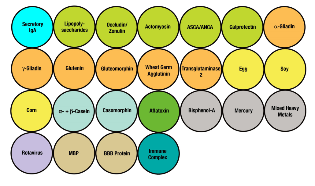
Secretory Immnuglobulin A (SIgA) is the first antibody isotype that appears in the bodily secretions being detectable at 2 to 4 weeks of age. It is composed of two IgA molecules (dimeric IgA), a joining protein (J), and a secretory component (SC).

The dimeric IgA-J chain complex is produced by B lymphocytes in the submucosal tissues.

The SC is produced by epithelial cells and acts as a receptor for dimeric IgA.

SIgA is the main immunoglobulin found in mucous secretions, including tears, saliva, sweat, colostrum, and secretions from the genitourinary tract, GI tract, prostate and respiratory epithelium. It is the most abundant class of antibodies found in the intestine. It promotes clearance of microbial pathogens and antigens from intestinal lumen through a process of entrapment called “immune exclusion.” SIgA uses this process to prevent the binding of these invaders to the epithelial cell receptors and entrapping them in the upper and lower layers of mucus, thus facilitating their removal through agglutination and GI secretion (see Figure 2).

SIgA also plays a role in maintaining mucosal immune homeostasis through the induction of oral tolerance by promoting the retrotransport of food antigens, bacterial toxins and more across the intestinal epithelia to dendritic cells. This is for the purpose of downregulating the proinflammatory responses that are normally associated with the uptake of these potentially inflammatory and allergenic antigens.

However, when the mucosal immune system is overwhelmed with food antigens, enteric toxins, pathogenic microorganisms and chemical toxicants, the mucosal immune system may lose its homeostasis. This loss of intestinal homeostasis can result in a much higher level of antigens across the epithelium, where the gut-associated lymphoid tissue can transfer the antigens into the circulation.

The presence of these unwanted antigens in the blood results in an immune response consisting of the production of IgA and IgM antibodies against them. These IgA/IgM antibodies acquire their secretory components, then find their way into bodily secretions, including saliva. This phenomenon is called mucosal leakiness to an antigen, which is an early event in the induction of autoimmunity in the gut and beyond.

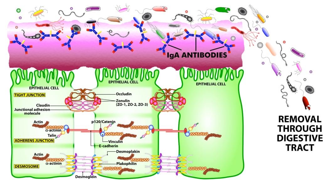
Figure 2. The immune system’s defensive wall. The mucous membrane or mucosa covers various cavities and internal organs of the human body, including the gastrointestinal tract. It is composed of a mucus layer that contains secretory IgA antibodies, the immune system’s first line of defense. SIgA antibodies catch antigenic invaders through the process of immune exclusion before they can penetrate epithelial cells, and eliminate the unwanted antigens through the digestive system.

SIgA has a wide range of critical functions in the body. These include the following:

  • As the first line of defense, SIgA provides a protective wall against harmful viruses, microbes and their antigens.
  • Under neuroinflammatory conditions, SIgA secreting cells travel from the gut to the brain with the help of regulatory cytokines such as interleukin-10 to attenuate neuroinflammation.SIgA can neutralize bacterial toxins and the neoantigens formed by toxic chemicals, such as when heavy metals bind to human tissue antigens.
  • SIgA can bind to viruses, preventing their Infection, Replication and Spread into the epithelial cells.
  • SIgA helps limit the presence of food antigens in the epithelium, thus preventing the initiation of immune response against food and other antigens.
  • By binding to a wide range of commensal bacteria, SIgA supports bacterial symbiosis by facilitating or modifying bacterial networks, or by removing them from the GI system.Despite these protective functions of SigA, in the presence of dysregulated gut immune function, breakdown in the oral tolerance mechanism, and large amounts of undigested food or other antigens, the immune system may trigger the overproduction of antigen-specific SIgA, which may contribute towards inflammation and autoimmunity in the gut and beyond.
  • The production of high levels of IgA antibodies in saliva against high number or volumes of antigens results in the formation of IgA-immune complexes.

The formation of these food, bacterial toxin, and neoantigen immune complexes may contribute to the failure of oral tolerance, the activation of inflammatory cascade, and the unwanted penetration of food, bacterial toxins and neoantigens into the submucosa and circulation. In addition, immune complexes that are formed by the binding of food antigens, bacterial toxins (LPS, BCDT) or neoantigens to their specific antibodies can bind to a very specific IgA receptor called CD71 on the surface of epithelial cells. This binding of antigen-antibody formations, for example, gliadin-IgA complexes or LPS-IgA complexes, to the IgA receptor may promote the entry of gliadin, LPS or other peptides or antigens through transepithelial transport into the submucosa and into the blood.

The transport of intact peptides through the intestinal epithelium perpetuates inflammatory and immune responses against the penetrating antigens, resulting in the production of IgA and IgM in the saliva, as well as the possible production of IgG (or other antibody isotypes), if the intact antigens or peptides reach the blood.

Array 14 is a cost-effective, easy, and non-invasive method for measuring mucosal immune reaction to a range of exogenous and endogenous antigens.

The production of IgA and IgM antibodies against antigens in saliva may be an early stage in the induction of inflammation and autoimmunity. If the immune reactivity is not addressed and it may result in a breach of the intestinal barrier, followed by a systemic immune reaction. The inflammation of systemic autoantibodies contributes to the progression of autoimmune and neuroautoimmune reactivity.

Array 14 was developed based on the immunological occurrence that exposure to an antigen via the mouth and intestinal tract results first in the production of IgA antibodies in secretions. Repeated exposure to the same antigen(s) results in the production of IgG, IgM and IgA antibodies in the blood. The biomarkers in Array 14 were selected to give an invaluable picture of both likely antigens which may cause immune reactions and the areas they may effect. These include the following:

  • Commonly consumed foods such as wheat, lectins, milk, soy, corn and egg
  • Gut microbiota-associated toxins
  • Toxic chemicals that contaminate food and form neoantigens with food proteins, such as aflatoxin, bisphenol-A and heavy metals
  • Rotavirus, which is involved in GI inflammation and the induction of food immune reactivity and autoimmunity
  • Tight junction/structural proteins involved in leaky gut and inflammation
  • Crohn’s disease and ulcerative colitis biomarkers such as ASCA, ANCA and calprotectin
  • Enzymes such as transglutaminase-2 that are involved in celiac disease and non-celiac gluten sensitivity
  • Enteric nerve/neural antigens such as myelin basic protein and S100B (BBB) because enteric nerve cells can be damaged by inflammation in the gut. Similar to CNS astrocytes, enteric glia cells express S100B, which is responsible for the maintenance of inflammation in the gut.
  • Immune complexes can be formed by triggers such as bacterial antigens, viral antigens, mold antigens, xenobiotics, dietary components, and associated tissue antigens, which further contribute to the entry of antigens, immune complexes, or other inflammatory molecules into the submucosa, and then into circulation. Due to structural similarity between the intestinal barrier and blood-brain barrier (BBB), these IgA + IgM antibodies, immune complexes and inflammatory signals can also affect the integrity of the BBB resulting in autoimmunity against nervous system tissues, both in the gut and the brain.

ARRAY 14 – Mucosal Immune Reactivity Screen™

IgA + IgM in Oral Fluid against 25 Antignes


Total SIgA

Gut/Barriers related

  • Lipopolysaccharie
  • Occludin/Zonulin
  • Actomyosin
  • Rota virus

Condition related

  • ASCA/ANCA
  • Calprotectin

Gluten related

  • Native + Deamidated Alpha-Gliadin-33-mer
  • Gamma Gliadin 15-mer
  • Glutenin 22-mer
  • Gliadin 33-mer
  • Gluteomorphin
  • Wheat Germ Agglutinin
  • Transglutaminase-2

Other Foods

  • Egg
  • Soy
  • Corn
  • Alpha-Casein
  • Beta-Casein
  • Casomorphin

Chemicals

  • Bisphenol-A
  • Mixed Heavy Metals
  • Mercury

Immune Related

  • Total SIgA
  • Immune Complex

BBB and Neural Related

  • Blood-Brain Barrier Protein
  • Myelin Basic Protein

The biomarkers in Array 14 were developed to shed light on important clinical information present in the mucous membrane.

Individual biomarkers, a grouping of the biomarkers, or the totality of the biomarkers, each reveal a part of an invaluable clinical picture.

The individual biomarkers give details on reaction to a single antigen or specific location of damage.

Each grouping gives a bigger picture of antigenicity or potential of more structural damage, and the panel as a whole may give a systemic picture of the immune status.

These include identifying possible loss of oral/immune tolerance, poly reactivity, complete loss of SigA or suppressed immune system, etc.

Exploring these mucosal antibodies may help to understand the development of immune reactivity through its course of action in the system.

The following is a look at a few case results:

Case 1 – Presented with:

  • 4 year old female
  • digestive problems: Intolerance to wheat, dairy, corn, eggs, occasional diarrhea, bloating, gas, abdominal pain,
  • Stuffy nose
  • Dry skin
  • Occasional headache, fatigue

Results show that not only is Secretory IgA very low, but SIgA specific against 28 different antigens representing tight junction proteins, food, toxic chemicals and blood-barrier proteins were also very low.

Results indicate patient has mucosal immune deficiency, which means she has no protective wall

Case 2

  • Test results in this case show possible sources of reactivity, intestinal permeability and progression of immune reactivity to BBB.
  • Mucosal reaction to gluten, wheat lectin, corn and aflatoxin
  • Mucosal reactivity to BPA found in plastics
  • Potential bacterial endotoxemia
  • Potential Intestinal permeability
  • Potential Intestinal autoimmunity
  • Mucosal immune cross-reactivity with blood-brain barrier proteins
  • Mucosal immune reactivity –

Case 3

  • Presented with: A 54-year-old female dentist.
  • 5ft, 99 lbs.
  • Complained of constant GI bloating even in a fasting state
  • She was born in south east Asia, and was diagnosed with vitiligo when she was 3.
  • At the age of 16 she moved to the US.
  • She had a significant amount of stress when she was running her own private dental practice.
  • She had muscle pain in her neck and shoulder area, and worked with a chiropractor for spinal correction.
  • She had sinus congestion, waking up once per night but able to get back to sleep.

Test results showed that the patient’s gut was very inflamed, and that her body was making polyreactive neutralizing antibodies to calm down the inflammation, like sprinklers being turned on to put out a fire.

The patient was put on a diet that eliminated gluten, dairy, soy and egg.

She is currently eating a plant-based diet with some fish, and claims 75% improvement since changing her diet.

Her bowel movements are normal.

She now works only part-time, and she claims to have good energy and mental clarity.

Antigens tested

Clinical significance

What to consider

Total SIgA High SIgA – GI tract under fire, inflammation and autoimmunity Remove all positive reactive antigens from the patient’s lifestyle and support with anti-inflammatories
Low SIgA – Possible mucosal immune deficiency and autoimmunity Cow and camel colostrum, L-glutamine, glutathione, green tea, probiotics, fiber, exercise
Lipopolysaccharide Gut dysbiosis Probiotics, L-glutamine
Occludin/Zonulin, Actomyosin Early stage of autoimmune reactivity to intestinal tight junction proteins, leaky gut Probiotics, L-glutamine, glutathione, regulation of regulatory T cells by vitamins A, C, D and AhR, acetate, butyrate, propionate
ASCA/ANCA, Calprotectin Yeast infection, activation of neutrophils in the gut tissue, release of enzymes, inflammation and autoimmunity in the gut and beyond Probiotics, L-glutamine, glutathione, anti-inflammatories, Curcumin, green tea, vitamins A, C, D
a-gliadin, g-gliadin, Glutenin, Gluteomorphin, Wheat germ agglutinin Failure of oral tolerance, mucosal gluten reactivity, celiac disease, non-celiac gluten sensitivity, lack of digestive enzymes including DPPIV Gluten-free and dairy-free diet, digestive enzymes including DPPIV
Transglutaminase Early immune reactivity to the intestinal villi antigens and enzymes, possible celiac disease, non-celiac gluten sensitivity, autoimmunity Gluten-free and dairy-free diet, support regulatory T cells with vitamins A, C, D and AhR
a-,b-casein, Casomorphin Failure of oral tolerance and dairy immune reactivity Gluten-free and dairy-free diet, digestive enzymes including DPPIV
Corn, Soy, Egg Failure of oral tolerance, immune reaction to these food antigens, their cross-reactivity with human tissue Removal of offending foods from diet.
Aflatoxin Over-exposure to food containing aflatoxin, immune reaction to food proteins and aflatoxin Oral glutathione, change in dietary habits including organic diet
Bisphenol-A, Mercury, Mixed heavy metals Oral and mucosal exposure to plastic materials and heavy metals that bind to proteins in the GI tract Lifestyle modification, liquid in non-plastic bottles, particular attention to fish consumption, oral glutathione, detoxification
Rotavirus Chronic exposure to rotavirus and induction of inflammation, celiac disease, non-celiac gluten sensitivity Probiotics, anti-inflammatory factors, gluten-free diet, regulation of regulatory T cells by vitamins A, C, D and AhR
Myelin basic protein, BBB protein Inflammation in the gut, damage to enteric nerve and release of neuronal antigens, disturbance in gut motility, neuroautoimmunity Probiotics, glutathione, N-acetylcysteine, fiber, acetate, butyrate, propionate, green tea extract, minocycline, curcumin
Immune Complexes Excessive antigen-antibody reaction, activation of complement cascade, induction of leaky gut, entry of many food antigens into circulation Removal of Array 4 and Array 10 immunoreactive foods from diet, probiotics, L-glutamine, glutathione, fiber, acetate, butyrate, propionate, vitamins A, C, D

Abnormal results

Recommended Follow-Up Tests or Arrays

Low or High Secretory IgA Array 2, Array 3, Array 4, Array 5, Array 10
Positive LPS, Occludin/Zonulin, Actomyosin antibody Array 2, Array 3, Array 5, Array 20
Elevated ASCA/ANCA, Calprotectin antibody Array 2, Array 5, Array 22
Elevated antibodies to a-gliadin, g-gliadin, Glutenin, Gluteomorphin, Wheat germ agglutinin Array 2, Array 3
Transglutaminase Array 2, Array 3
Elevated antibodies to a-,b-casein, Casomorphin Array 4, Array 5
Elevated antibodies to Corn, Soy, Egg Array 2, Array 10, Array 20
Elevated antibodies to Aflatoxin, Bisphenol-A, Mercury, Mixed heavy metals Array 11
Elevated antibody to Rotavirus Array 2, Array 3, Array 12
Myelin basic protein, BBB protein Array 5, Array 20
Immune Complexes Different combinations of arrays

How to Order

(for healthcare professionals)

NO NEED FOR BLOOD DRAW!

The test is self-administered at home.
All that is needed is a Cyrex Oral Fluid Specimen Collection Kit which can be sent directly to patient.

Step 1

Create a patient test requisition at cyrexlabs.com

Step 2

Request an Oral Fluid Test Kit – To be shipped directly to the patient
(877) 772-9739 Option 1

Step 3

Patient follows instructions to collect saliva sample at home and ships to Cyrex prepaid via UPS

TO LEARN MORE ABOUT OTHER CYREX TESTS

Call (877) 772-9739, Option 1
or email [email protected]

JoinCyrex.com

©SIgATest 2020. All Rights Reserved